Cherry Angioma Removal: What Are They and How Can They Be Treated?
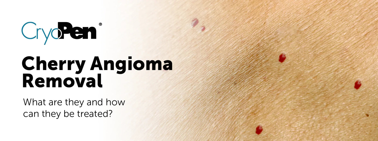

Cherry angiomas are very common, harmless red skin marks made up of tiny blood vessels. They often appear as small bright red dots or raised red bumps on the skin and are most frequently seen on the torso, arms, back and shoulders.
Although cherry angiomas are usually benign, many people choose to have them removed because they feel self-conscious, the lesion catches on clothing, or it bleeds when knocked or scratched.
In the UK, cherry angioma removal is usually carried out in a professional clinic using methods such as cryotherapy, laser treatment or electrocautery. CryoPen is one of the most recognised precision cryotherapy devices used by trained practitioners to treat benign skin lesions, including cherry angiomas.
CryoPen uses a controlled jet of nitrous oxide to freeze the unwanted tissue with accuracy, helping practitioners target the lesion while reducing unnecessary impact on the surrounding skin.
For patients considering treatment, the most important step is choosing the right clinic. Look for an authorised CryoPen clinic where practitioners have completed proper CryoPen training, often supplied through IvanMed, the exclusive CryoPen supplier and educator in the UK.
What Is A Cherry Angioma?
A cherry angioma, also known as a Campbell de Morgan spot, is a benign vascular lesion. It forms when small blood vessels cluster near the surface of the skin, creating a red, ruby-coloured or purple-red mark.
Cherry angiomas are not usually a medical concern. They can be flat or slightly raised, very small or several millimetres wide. They tend to become more common with age and may increase in number over time.
Most people seek removal for cosmetic reasons, although removal may also be considered if the angioma is irritated, catches on clothing, or bleeds when traumatised.
When Should A Red Skin Mark Be Checked First?
Even though cherry angiomas are usually harmless, any skin lesion should be assessed properly before cosmetic removal. You should seek medical advice before treatment if the lesion:
-
Changes quickly in size, shape or colour
-
Bleeds repeatedly without being knocked
-
Looks very different from your other skin marks
-
Becomes painful, ulcerated or persistently inflamed
-
Has an irregular border or unusual colouring
-
Is not clearly identifiable as a benign lesion
A trained CryoPen practitioner should not treat a lesion if they are not confident that it is benign. Our CryoPen treatment guidance advises practitioners not to proceed if they are unsure and to refer clients to a GP or suitable lesion-checking service where needed.
Cherry Angioma Removal Options In The Uk
There are several ways to remove cherry angiomas in a clinic. The best option depends on the size, depth, location, skin type, medical history and practitioner assessment.
Cryotherapy with CryoPen
Cryotherapy works by freezing the targeted lesion so the treated tissue breaks down and naturally heals away. CryoPen is a pen-like cryotherapy device that delivers nitrous oxide directly to benign skin lesions.
CryoPen is designed for precision. Unlike some other freezing methods, it allows trained practitioners to focus the treatment on the lesion itself. This makes it a popular option for many benign cosmetic lesions, including cherry angiomas, skin tags, warts, verrucas, sun spots and seborrhoeic keratoses.
For professionals, CryoPen is valuable because it offers controlled application, different applicator options and treatment flexibility. The CryoPen is a portable, pen-like device that uses disposable nitrous oxide cartridges and allows practitioners to treat benign lesions with pinpoint accuracy.
What to expect with CryoPen treatment:
-
The treatment is usually quick, often lasting seconds per lesion
-
The practitioner will assess the lesion, skin type, size and depth before treatment
-
You may feel cold, stinging or a short sharp sensation
-
The treated area may become red, swollen, darker, blistered or scabbed as it heals
-
More than one treatment may be needed for some lesions
Practitioner technique matters. We advise practitioners to assess the lesion, choose the correct applicator, determine the treatment time, allow the area to thaw, repeat the freeze cycle where appropriate, and schedule follow-up after two to four weeks.
Laser treatment
Laser treatment targets the blood vessels inside the cherry angioma. It is commonly used for vascular lesions and may be suitable when treating multiple angiomas or when the practitioner feels laser is the better option for the area, skin type or lesion presentation.
Laser treatment can be effective, but patients should still expect a consultation, skin assessment, aftercare advice and a discussion about possible side effects such as redness, swelling, pigmentation changes or temporary sensitivity.
Electrocautery
Electrocautery uses heat to cauterise and remove the lesion. It can be effective for raised or easily traumatised angiomas, but correct technique is essential to reduce the risk of scarring, pigmentation change and delayed healing.
As with any lesion removal treatment, electrocautery should only be carried out after the lesion has been assessed and confirmed as suitable for cosmetic removal.
How Many Sessions Are Needed For Cherry Angioma Removal?
Some cherry angiomas respond after one session. Others may need more than one treatment depending on the size, depth, location and individual healing response.
With CryoPen, most lesions can respond to a single treatment, although deeper lesions may require further sessions.
Your practitioner should explain this during consultation so you know what to expect before booking treatment.
Healing Timeline After Cherry Angioma Removal
Healing varies from person to person. It also depends on the treatment method, lesion depth, body area and aftercare.
After CryoPen treatment, the area may go through the following stages:
First few hours: Mild stinging, redness and slight swelling are common.
First few days: The lesion may darken, blister or begin to form a crust.
One to three weeks: A scab or crust may develop and fall away naturally.
Two to six weeks: The skin continues to heal and remodel. Some areas, especially on the body or lower limbs, may take longer.
Healing typically occurs within two to six weeks depending on the freeze time and treatment location. If the treated area has not healed after six weeks, the patient should contact their treatment provider.
Aftercare After Cherry Angioma Removal
Good aftercare helps reduce the risk of infection, delayed healing, scarring and pigmentation problems. Your practitioner should give you specific advice based on the treatment area.
General aftercare principles include:
-
Do not pick, scratch or pull off the scab
-
Keep the area protected from friction and rubbing
-
Avoid applying products unless advised by your practitioner
-
Keep the area as dry as possible during the early healing stage
-
Avoid swimming, saunas, steam rooms, sunbeds and excessive sweating while advised
-
Use SPF once the area has healed, especially if there is any pigmentation change
-
Contact your clinic if the area becomes increasingly painful, hot, swollen, red, weepy or produces pus
If a blister forms, do not deliberately pop it. If it opens accidentally, follow your practitioner’s instructions and keep the area clean..
Will Cherry Angioma Removal Leave A Scar Or White Mark?
Any skin procedure can leave a mark, including cryotherapy, laser or electrocautery. However, many patients heal well when the correct treatment is chosen and aftercare is followed.
The main risks to discuss before treatment are:
Pigment change: The treated area may heal lighter or darker than the surrounding skin. This can last a few months and may occasionally be longer lasting.
Hypopigmentation in darker skin tones: Darker skin types have a higher risk of pigment loss after cryotherapy because melanocytes are sensitive to cold injury.
Scarring: Scarring is possible with any lesion removal method, especially if the area is over-treated, picked, infected or slow to heal.
CryoPen consultation documents list pigment changes, rare nerve damage, blistering and hair follicle damage in hair-bearing areas as possible risks and side effects.
A good clinic will explain these risks clearly before treatment, rather than promising a mark-free result.
Does Cherry Angioma Removal Hurt?
Most people find cherry angioma removal tolerable. With CryoPen, anaesthetic is often not required because the extreme cold has a temporary numbing effect during treatment.
You may feel a cold, sharp, stinging or aching sensation during the freeze. Some residual stinging can continue for a short time afterwards. Little or no discomfort is usually felt during the first few seconds when healthy tissue is avoided, although discomfort may increase once the cold reaches the depth of the lesion.
Why Professionals Use Cryopen For Benign Lesion Removal
For clinics, CryoPen can be a useful addition to professional lesion removal services because it offers fast treatment times, precision and a wide range of benign lesion indications.
CryoPen is used for benign skin lesions such as cherry angiomas, skin tags, warts, verrucas, seborrhoeic keratoses and solar lentigines. It delivers nitrous oxide directly to the targeted tissue, allowing the practitioner to treat with control and accuracy.
For professionals, the quality of training is just as important as the device itself. A trained practitioner should understand lesion recognition, contraindications, consultation, treatment timing, freeze-thaw technique, aftercare, complication management and when to refer.
IvanMed’s CryoPen training materials highlight the importance of consultation, lesion analysis, photographic documentation, contraindication checking and clear patient expectations before treatment.
Choosing A Cryopen Clinic In The UK
If you are considering cherry angioma removal, look for an authorised CryoPen clinic rather than choosing on price alone.
A good clinic should offer:
-
A proper consultation before treatment
-
A clear lesion-checking process
-
Honest advice if the mark needs medical assessment first
-
Trained CryoPen practitioners
-
Clear aftercare instructions
-
Realistic expectations about healing and pigmentation risk
-
A follow-up plan if another session is needed
Many authorised CryoPen clinics in the UK are trained through IvanMed, the official CryoPen supplier and training provider. Choosing a clinic with proper CryoPen training gives you more confidence that the practitioner understands safe treatment technique, benign lesion suitability and post-treatment care.
If you are unsure whether your red mark is a cherry angioma, book a skin assessment first. Cosmetic removal should only be considered once the lesion has been identified as benign.
FAQs
Can I Remove A Cherry Angioma At Home?
No, home removal is not recommended. Trying to cut, burn, freeze or pick off a cherry angioma at home can cause bleeding, infection, scarring and delayed healing. It can also delay proper assessment if the lesion is not actually a cherry angioma.
How Long Does Cryopen Treatment Take?
CryoPen treatment is usually quick, often lasting seconds per lesion. The exact timing depends on the size, depth, location and practitioner technique.
What Happens Straight After Cryopen Cherry Angioma Removal?
The area may look red, slightly swollen or darker. Mild stinging is common. Over the next few days, a blister, scab or crust may form as part of the normal healing process.
Can Cherry Angiomas Come Back After Removal?
A successfully treated cherry angioma is often removed permanently, but new cherry angiomas can appear elsewhere over time. This does not usually mean the treatment has failed.
Is Cryopen Suitable For All Skin Types?
CryoPen may be used on different skin types, but darker skin tones need careful risk assessment because of the higher risk of pigment change. Your practitioner should discuss this before treatment.
Do I Need A Medical Appointment Before Cherry Angioma Removal?
If the lesion is clearly benign and the practitioner is trained to assess suitability, treatment may be carried out in the clinic. If there is any doubt about the diagnosis, you should seek medical assessment before cosmetic removal.
Conclusion
Cherry angiomas are common, benign red skin lesions, but they can be frustrating if they catch on clothing, bleed when knocked, or affect confidence. In the UK, professional treatment options include CryoPen cryotherapy, laser and electrocautery.
CryoPen is a precision cryotherapy device used by trained practitioners to treat benign skin lesions, including cherry angiomas. It offers fast treatment, controlled application and targeted freezing when used correctly.
The safest approach is to choose an authorised CryoPen clinic with properly trained practitioners, ideally trained through IvanMed. A professional clinic should assess the lesion first, explain the risks, provide clear aftercare and advise medical referral if there is any uncertainty about what the lesion is.